Mastoiditis can result from untreated middle ear infections. Mastoiditis: Infection of the mastoid bone, just behind the ear.Hearing loss, vertigo, and tinnitus can be symptoms. Acoustic neuroma: A noncancerous tumor that grows on the nerve traveling from the ear to the brain.The small hole usually heals within a few weeks. Ruptured eardrum: Very loud noises, sudden changes in air pressure, infection, or foreign objects can tear the eardrum.The eardrum’s reduced vibrations impair hearing. Cerumen ( ear wax) impaction: Ear wax may block the ear canal and adhere to the eardrum.

Usually this is due to damage from noise exposure, or from aging. Tinnitus: Ringing in one or both ears.Vertigo, tinnitus, hearing loss, and pain are common symptoms. Meniere’s disease: A condition in which the inner ear on one side malfunctions.
AUDITORY CANAL FUNCTION SKIN
Sudden cases are usually infections chronic otitis is often a skin condition (dermatitis).
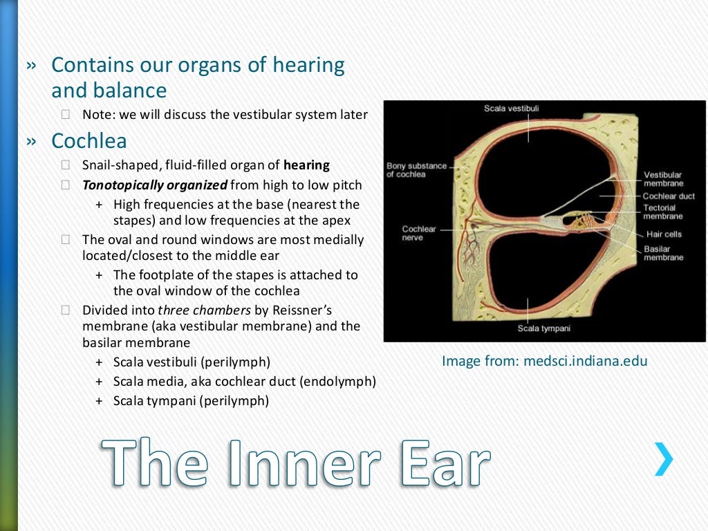
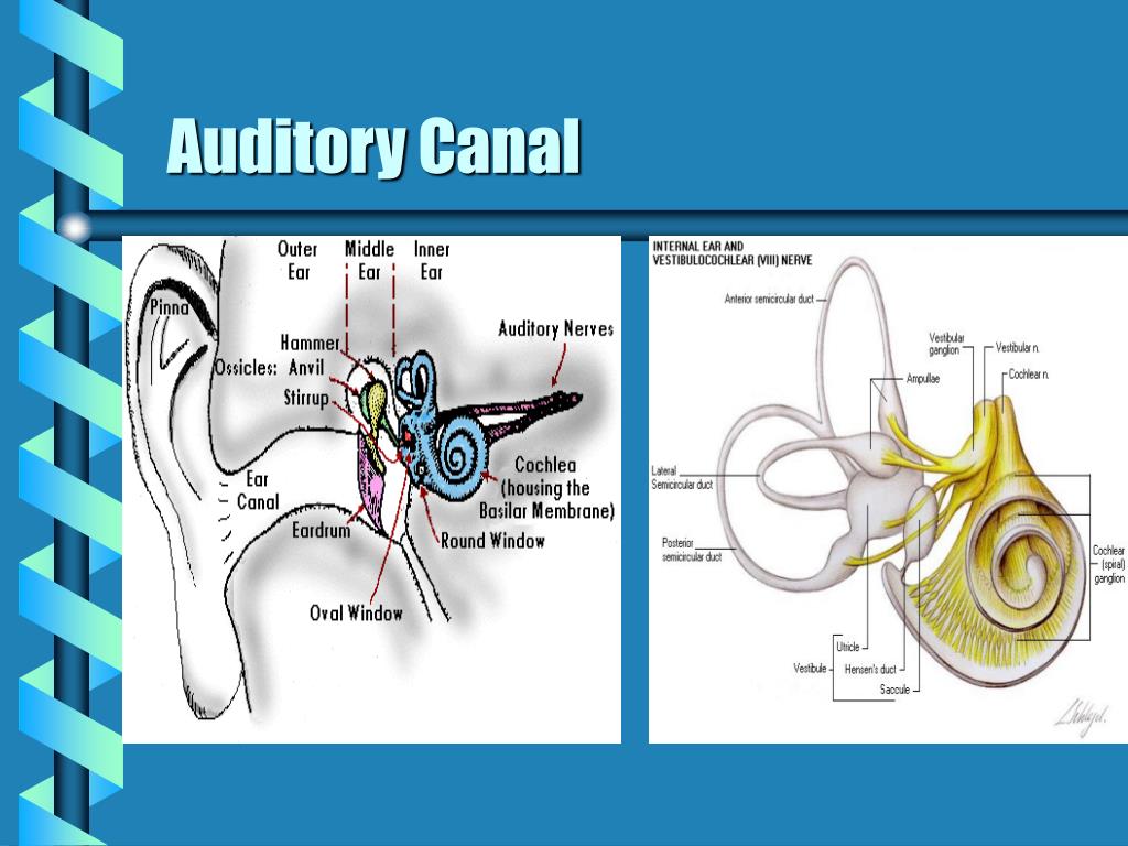
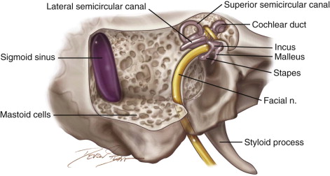
Swimmer’s ear (Otitis externa): Inflammation or infection of the outer ear (pinna and ear canal). Otitis media (middle ear inflammation): Inflammation or infection of the middle ear (behind the eardrum). Some of these are serious, some are not serious. Earache: Pain in the ear can have many causes. The eustachian (auditory) tube drains fluid from the middle ear into the throat (pharynx) behind the nose. They send information on balance and head position to the brain. The fluid-filled semicircular canals (labyrinth) attach to the cochlea and nerves in the inner ear. The spiral-shaped cochlea is part of the inner ear it transforms sound into nerve impulses that travel to the brain. Sound causes the eardrum and its tiny attached bones in the middle portion of the ear to vibrate, and the vibrations are conducted to the nearby cochlea. Sound funnels through the pinna into the external auditory canal, a short tube that ends at the eardrum (tympanic membrane). The outer ear is called the pinna and is made of ridged cartilage covered by skin. Conditions that affect the malleus often impact the ability to hear.The ear has external, middle, and inner portions. The malleus functions with the other bones to transmit vibrations from the eardrum to the inner ear. The malleus, also known as the “hammer” or “mallet,” is the largest of three small bones in the middle ear. Stirrup (stapes) - attached to the membrane-covered opening that connects the middle ear with the inner ear (oval window) Anvil (incus) - in the middle of the chain of bones. The middle ear contains three tiny bones: Hammer (malleus) - attached to the eardrum. The internal auditory canal (IAC), also referred to as the internal acoustic meatus lies in the temporal bone and exists between the inner ear and posterior cranial fossa. From that station, neural impulses travel to the brain – specifically the temporal lobe where sound is attached meaning and we HEAR. What does the auditory nerve do?Īuditory nervous system: The auditory nerve runs from the cochlea to a station in the brainstem (known as nucleus). The lateral surface of the tympanic membrane, the external auditory canal, and the external acoustic meatus are all innervated by nervus intermedius (a branch of CN VII), the auriculotemporal nerve (CN V3), and the auricular branch of the vagus nerve. What Innervates the external auditory canal? The fissures of Santorini are natural openings within the anterior cartilage in the lateral aspect of the canal that may allow infectious or neoplastic processes to extend from the parotid gland to the canal or the opposite scenario (Fig. 
The external acoustic meatus is directed in a horizontal oblique, medial and slightly anterior direction. What is the direction of external auditory canal?

Where does the external auditory canal end? The ear canal – the auditory canal The external auditory canal’s function is to transmit sound from the pinna to the eardrum.


 0 kommentar(er)
